Characterizing Planar SERS Substrates: Unraveling the Link between Physical Characteristics and Performance Metrics
Feb 2024
Mehdi Feizpour, Qing Liu, Tom Van der Donck, Hugo Thienpont, Wendy Meulebroeck and Heidi Ottevaere
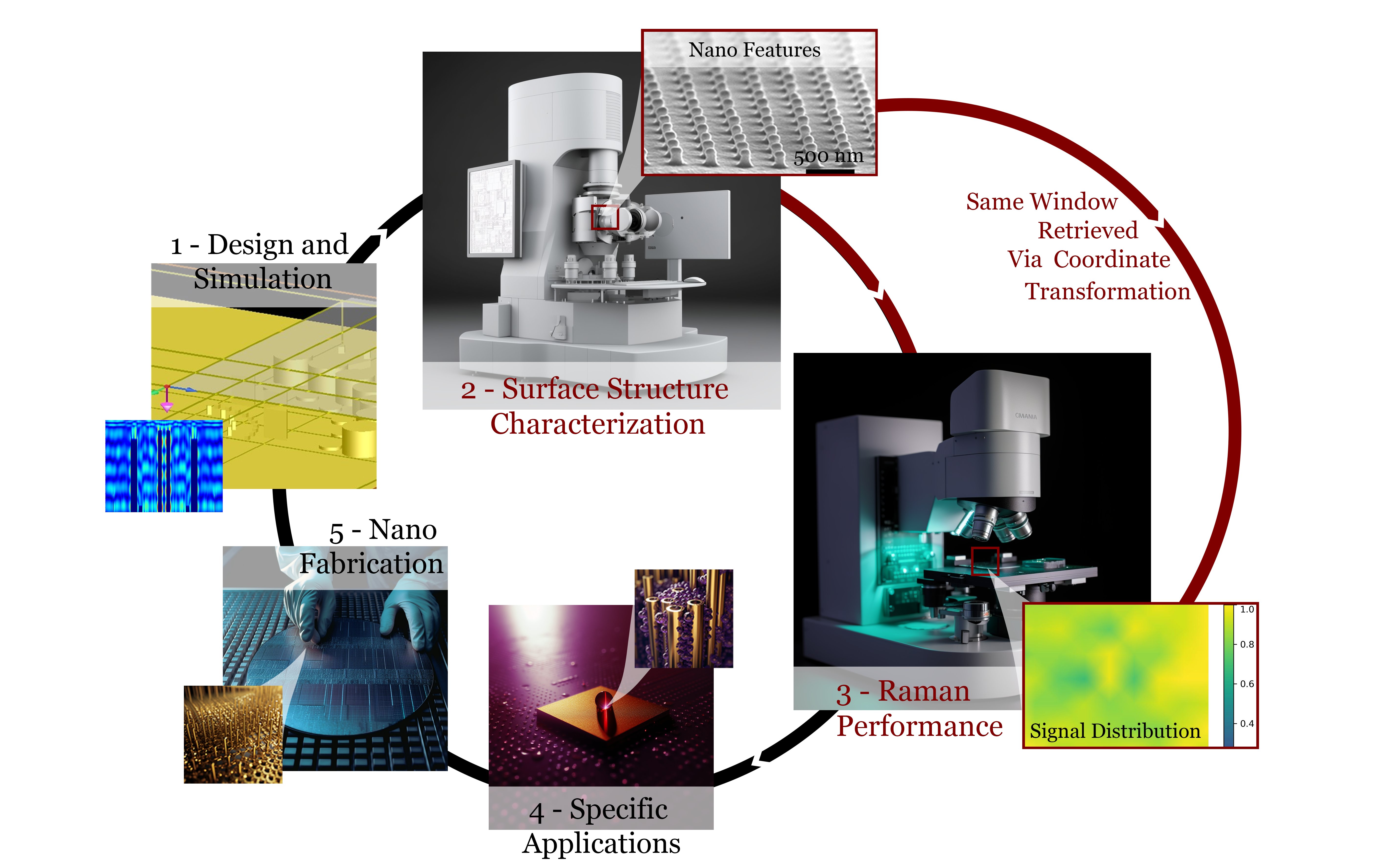
Surface-enhanced Raman spectroscopy (SERS) is a powerful optical sensing technique used in various applications, including medicine, microbiology, and environmental analysis. Planar SERS substrates are of particular interest due to their ease of integration in lab-on-chips and better reproducibility compared to colloidal SERS. The performance of these SERS substrates is quantified using metrics such as enhancement factor, sensitivity, and reproducibility. However, there is yet to be a consensus on how to practically compare and interpret such metrics in publications and experiments. These performance metrics are strongly influenced by the nanostructures’ material, architecture, element sizes, as well as the circumstances surrounding the experiments. Understanding the effect of these characteristics on the SERS substrates’ performance could not only enable a better performance but also direct their development for different applications. Thus, we developed a planar SERS-substrate characterization protocol to explore the correlation between the nanostructures’ physical characteristics and the performance metrics through coordinate-transformed spectroscopic measurements over structure-characterized areas. Seven commercial SERS substrates, with various surface architectures fabricated using different fabrication technologies, were studied using this benchmarking protocol. The results demonstrated how this protocol can indicate a SERS substrate’s suitability for a specific application, thus, guiding the substrate’s further adaptations or development.
Classification of hemoglobin fractions in the liquid state using Raman spectroscopy combined with machine learning
November 2023
Sara Abbasi, Mehdi Feizpour, Ilse Weets, Qing Liu, Hugo Thienpont, Francesco Ferranti, Heidi Ottevaere

Identifying hemoglobinopathies is important for the clinical management of many diseases. One of the common techniques to screen hemoglobinopathies is through high-performance liquid chromatography separation followed by UV–VIS detection. Although UV–VIS can quantify the hemoglobin fractions, it is unable to identify them. Here, we use Raman microscopy to generate fingerprint spectra of hemoglobin fractions based on which the fractions can be identified. Five different hemoglobin types are investigated in their liquid state: HbA0, HbS, HbF, HbA1c, and HbA2. Machine learning models based on support vector machines and fully-connected neural networks are optimized to classify these fractions achieving 98.2 ± 0.1% and 98.5 ± 0.3% test F1-score, respectively. In addition, the test accuracy of these two models are 98.2 ± 0.1% and 98.5 ± 0.3%, respectively. Our approach demonstrates the potential of Raman spectroscopy as an identification module in combination with high-performance liquid chromatography. Moreover, this detection approach can be easily miniaturized and integrated with microfluidics.
Development of two-photon polymerized (2PP) periodic nanostructures for label-free SERS biosensing
In Review
Tatevik Chalyan, Mehdi Feizpour, Koen Vanmol, Núria Solerdelcoll, Gen Takebe, Hugo Thienpont, Heidi Ottevaere, Qing Liu

Surface-enhanced Raman spectroscopy (SERS) has proven its powerful ability to precisely characterize a biological substance down to a single molecule level without the use of a ligand or a specific biofunctionalization. A variety of nanofabrication technologies enable SERS substrate prototyping and mass manufacturing. In this work, SERS substrate simulation, fabrication, and prototyping based on two-photon polymerization (2PP) are discussed. Developed substrates show up to 106 Raman signal enhancement, comparable to commercial substrates. Moreover, the rapid prototyping of SERS substrates based on 2PP takes from less than a minute to 2 hours depending on the fabrication approach and aspect ratio requirements. The process is well-controlled and reproducible for reaching a uniform distribution of nanostructure arrays. Developed SERS substrates can be used for a broad range of applications and characterization of different molecules.
Conference Proceedings

Analyzing SERS reproducibility and performance: the role of illumination area
Mehdi Feizpour, Qing Liu, Hugo Thienpont, Wendy Meulebroeck, Heidi Ottevaere
In this work, the role of the beam spot in the SERS performance of a planar substrate is discussed. The planar SERS substrates’ feature sizes define their application and limitations.
Fig: Maximum normalized EF maps of the 3 SEM measured windows of (a1-a3) PiCO Au, (b1-b3) ATO ID Ag, (c1-c3) Hamamatsu Au, (d1-d3) SERSitive Ag, (e1-e3) Silmeco Au for the 1636 cm-1 BPE peak and 20 μM concentration. The bar plot, (f), shows the absolute maximum EF averaged over the 3 maps. The error bars show the map-to-map variation of the maximum EF. The vertical axis has a factor of 1000.

Automatic optimization of spectral classifiers’ hyperparameters for pathogen identification through evolutionary techniques
Mehdi Feizpour, Sara Abbasi, Qing Liu, Thomas Demuyser, Hugo Thienpont, Wendy Meulebroeck, Heidi Ottevaere
The hyperparameters of a selection of classifiers were optimized using a genetic algorithm. The target data was the SERS spectra of CSF pathogens.
Fig: Optimization curve of the SVM classifier with a random initialization.

FDTD modeling of 2PP-fabricated SERS substrates: design, simulation and hotspot analysis
Qing Liu, Tatevik Chalyan, Mehdi Feizpour, Heidi Ottevaere
Finite-Difference Time-Domain (FDTD) simulations of two-photon polymerization (2PP) printed pillars.
Fig: (a) Height and pitch sweeping of the 250 nm voxel arrays with 25 nm gold coating. (b) The electric field amplitude of voxel arrays with a pitch of 560 nm and heights of 200nm, with a maximum amplitude of 5.

Two-Photon Polymerized nanopillars for Surface-Enhanced Raman spectroscopy
Tatevik Chalyan, Mehdi Feizpour, Heidi Ottevaere
Pushing the limits of two-photon polymerization (2PP) prints for custom SERS prototyping.
Fig: AFM image of a homogeneously fabricated SERS with P=15 mW; D=300 nm; Pitch=360/380 nm; h=1 µm.

Advancing Hemoglobinopathy Screening with Raman Spectroscopy and Machine Learning
Sara Abbasi, Mehdi Feizpour, Qing Liu, Ilse Weets, Hugo Thienpont, Francesco Ferranti, Heidi Ottevaere
Raman spectroscopy combined with machine learning shows potential for hemoglobinopathy screening.
Fig: ROC curves and confusion matrices of the a) PCA/SVM classifier and b) PCA/NN classifier. The area under the curve for each class is given in the plot’s legends.

Two-Photon Polymerization based fabrication of SERS substrates for biosensing applications
Tatevik Chalyan, Qing Liu, Mehdi Feizpour, Hugo Thienpont, and Heidi Ottevaere
The design and fabrication of Surface-Enhanced Raman Spectroscopy substrates based on two-photon polymerization of photoresist with direct laser writing.
Fig: (a) 3D AFM image of the gold coated SERS substrate. (b) SERS spectrum of CV of 5.5µM.
