There are some upcoming articles. You can track them below as “Under Review”.
SERS Substrate Fabrication via Rapid Triboelectrification-Driven Self-Assembly of Close-Packed Colloidal Monolayers
Patent Pending, Under Review in Small Methods
Mehdi Feizpour, Ignaas Jimidar, Gert Desmet, Heidi Ottevaere
Bacterial Identification in SERS-Integrated Microfluidics Using CNN-Driven 2D Classification of 1D Spectra
Dec 2025
Mehdi Feizpour, Halewijn Van den Bosche, Lilit Melikyan, Thomas Demuyser, Piet Cools, Hugo Thienpont, Tatevik Sarukhanyan, Heidi Ottevaere
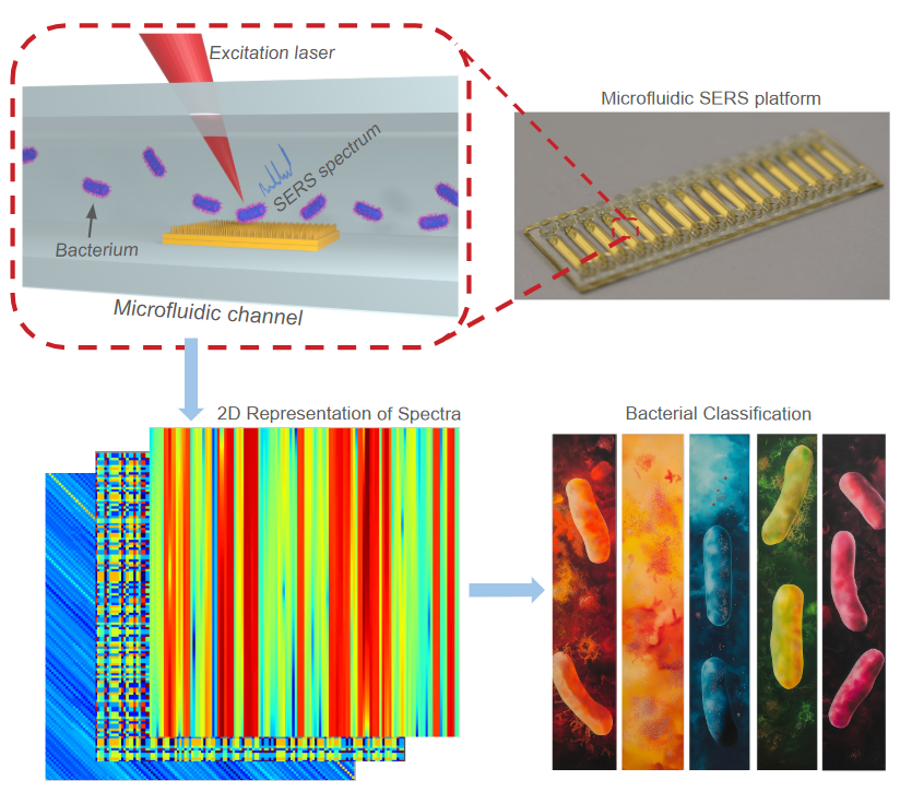
Bacterial sensing involves complex and variable samples that require advanced handling and analytical methods. To address these challenges, machine learning—especially deep learning—and SERS-based microfluidics have shown great promise. While previous studies have majorly focused on 1D spectral classification, the use of 2D representations of SERS spectra has not yet been explored, particularly for on-chip bacterial identification. In this work, we introduce a novel framework that combines SERS-enabled microfluidics with optimized 2D convolutional neural networks (2D-CNNs) for bacterial classification. SERS integration inside microfluidic chips was achieved through direct laser writing, enabling custom active areas and efficient on-chip measurements. We systematically evaluated nine distinct 1D-to-2D spectral transformations, with spectrogram and continuous wavelet transform yielding test accuracies of 99% and 97%, respectively, on controlled datasets. Using transfer learning, we achieved 100% accuracy on the on-chip dataset, demonstrating the model’s adaptability to new data. In contrast, other transformations, like pairwise distance and autocorrelation, performed below 93%, indicating their limited ability to capture subtle spectral features. This framework offers high sample control, parallelization, and the potential for expanding the bacteria database, making it ideal for low-data-volume situations such as rare infections. Further development and testing across strains, environments, and practical challenges can further improve our approach’s reliability for real-world diagnostics.
Direct Laser Writing of Curved SERS for Light-guided Enhancement
Nov 2024
Mehdi Feizpour, Sara Abbasi, Hugo Thienpont, Heidi Ottevaere
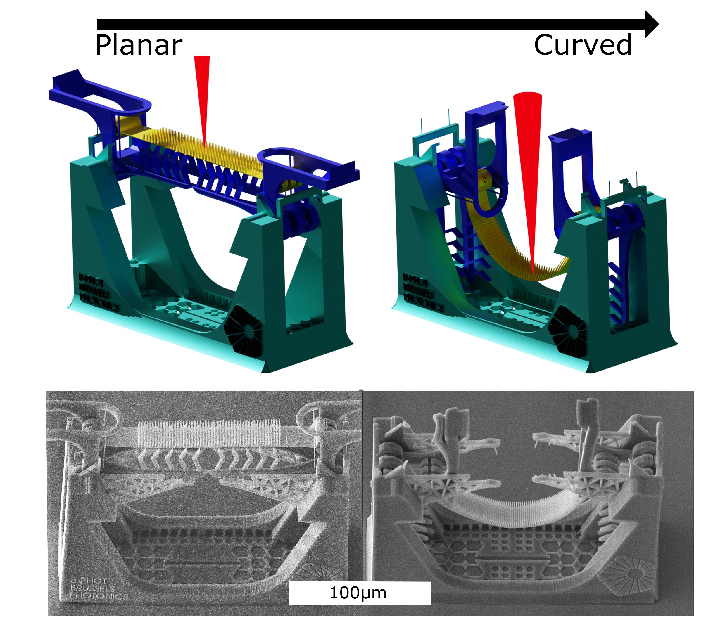
Flexible SERS substrates, typically used on irregular surfaces, have unexplored optomechanical effects that could enhance performance. We developed a micromechanically bending structure, i.e. microbender, to study how bending affects SERS substrates’ performance. Optical simulations of nanopillar arrays on micro-curved surfaces showed a 25–134 % improvement in mean-field localization at the pillar tips for arrays with pillar diameters of 0.4–1 m, pitches of 0.5–0.8 μ m tall pillars (0.5 μ m, and heights of 0.5–4.5 μ m. Raman measurements showed that SERS intensity increases when in a curved state due to enhanced light scattering, guiding, and field localization. A curved array of 6 μ arrays with shorter 0.8 μ m diameter, 1 m pillars (0.2 μ m spacing) produced Raman intensities similar to dense single-voxel μ m diameter, 0.2 μ m gaps) in the planar state. This finding suggests that low resolution fabrication technologies can produce curved SERS substrates with similar performance to high resolution planar SERS, offering an alternative to the current “smaller is better” trend through post- fabrication manipulation.
Characterizing Planar SERS Substrates: Unraveling the Link between Physical Characteristics and Performance Metrics
Feb 2024
Mehdi Feizpour, Qing Liu, Tom Van der Donck, Hugo Thienpont, Wendy Meulebroeck and Heidi Ottevaere
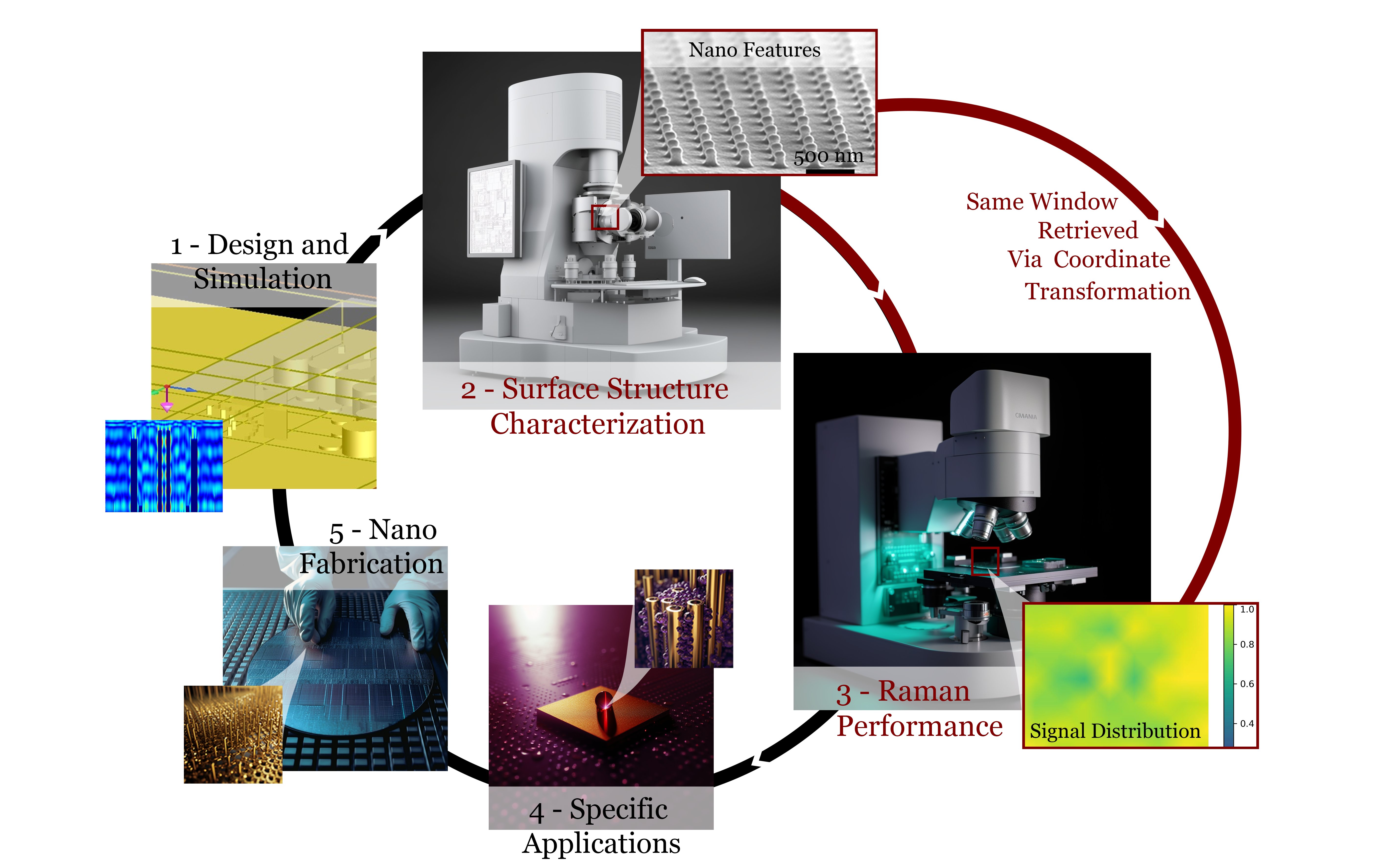
Surface-enhanced Raman spectroscopy (SERS) is a powerful optical sensing technique used in various applications, including medicine, microbiology, and environmental analysis. Planar SERS substrates are of particular interest due to their ease of integration in lab-on-chips and better reproducibility compared to colloidal SERS. The performance of these SERS substrates is quantified using metrics such as enhancement factor, sensitivity, and reproducibility. However, there is yet to be a consensus on how to practically compare and interpret such metrics in publications and experiments. These performance metrics are strongly influenced by the nanostructures’ material, architecture, element sizes, as well as the circumstances surrounding the experiments. Understanding the effect of these characteristics on the SERS substrates’ performance could not only enable a better performance but also direct their development for different applications. Thus, we developed a planar SERS-substrate characterization protocol to explore the correlation between the nanostructures’ physical characteristics and the performance metrics through coordinate-transformed spectroscopic measurements over structure-characterized areas. Seven commercial SERS substrates, with various surface architectures fabricated using different fabrication technologies, were studied using this benchmarking protocol. The results demonstrated how this protocol can indicate a SERS substrate’s suitability for a specific application, thus, guiding the substrate’s further adaptations or development.
Toward nanofabrication of SERS substrates with two-photon polymerization
Dec 2024
Tatevik Chalyan, Mehdi Feizpour, Qing Liu, Koen Vanmol, Núria Solerdelcoll, Gen Takebe, Hugo Thienpont, Heidi Ottevaere
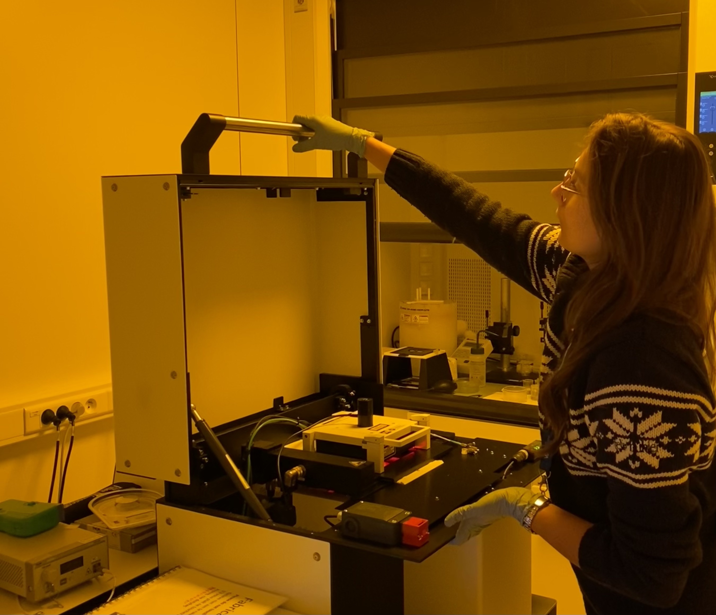
Surface-enhanced Raman spectroscopy (SERS) has shown its ability to characterize biological substances down to a single-molecule level without a specific biorecognition mechanism. Various nanofabrication technologies enable SERS substrate prototyping and mass manufacturing. This study reports a complete cycle of design, fabrication, prototyping, and metrology of SERS substrates based on two-photon polymerization (2PP). Highly controllable direct laser writing allows the fabrication of individual nanopillars with up to an aspect ratio of 4. The developed SERS substrates show up to 106 Raman signal enhancement, comparable to commercial substrates. Moreover, the rapid prototyping of the 2PP-printed SERS substrates takes from a minute to less than 2 hours, depending upon the nano-printing approach and aspect ratio requirements. The process is well-controlled and reproducible for achieving a uniform distribution of nanostructure arrays, allowing the SERS substrates to be used for a broad range of applications and the characterization of different molecules.
Classification of hemoglobin fractions in the liquid state using Raman spectroscopy combined with machine learning
November 2023
Sara Abbasi, Mehdi Feizpour, Ilse Weets, Qing Liu, Hugo Thienpont, Francesco Ferranti, Heidi Ottevaere

Identifying hemoglobinopathies is important for the clinical management of many diseases. One of the common techniques to screen hemoglobinopathies is through high-performance liquid chromatography separation followed by UV–VIS detection. Although UV–VIS can quantify the hemoglobin fractions, it is unable to identify them. Here, we use Raman microscopy to generate fingerprint spectra of hemoglobin fractions based on which the fractions can be identified. Five different hemoglobin types are investigated in their liquid state: HbA0, HbS, HbF, HbA1c, and HbA2. Machine learning models based on support vector machines and fully-connected neural networks are optimized to classify these fractions achieving 98.2 ± 0.1% and 98.5 ± 0.3% test F1-score, respectively. In addition, the test accuracy of these two models are 98.2 ± 0.1% and 98.5 ± 0.3%, respectively. Our approach demonstrates the potential of Raman spectroscopy as an identification module in combination with high-performance liquid chromatography. Moreover, this detection approach can be easily miniaturized and integrated with microfluidics.
